Loading...
Loading...
Toggle navigation
Products
Products
Newborn Screening
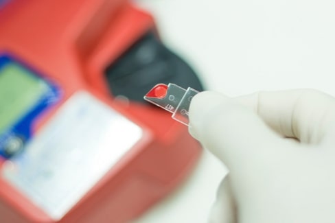
Point of Care Test
Hand hygiene products
Surface Disinfectants
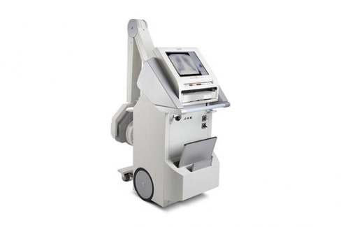
Computerised Radiography
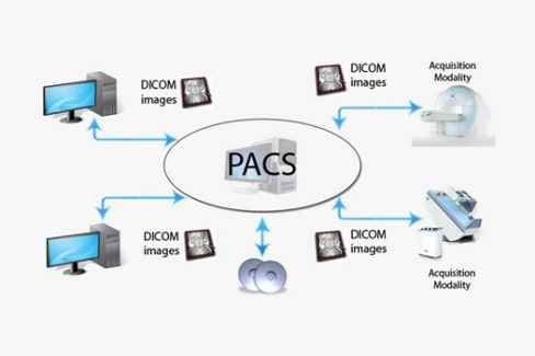
PACS Solutions
Radiology Printing Solutions
Veterinary Radiography
In-Vitro Diagnostics
Covid-19
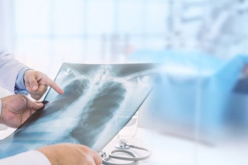
Radiography & C-Arm Systems
Mammography
Ultrasound
HPLC
Radiation Protection
ICU Solutions
Clinical Chemistry
Hematology
Immunology
Infectious Disease
Gastroenterology
Radiology Accessories
View all
Specialties
Specialties
Consumables
Gynecology
Immunology
Laboratory
Mammography
Radiology
Turnkey projects
View all
Media
About
Careers
Reach us
Global
China
Finland
Germany
India
Japan
Nepal
Oman
Sri Lanka
Turkey
UAE
follow us
Facebook
Twitter
linkedin
Instagram
youtube
Home
Products
Computerised Radiography
Covid-19
Hand hygiene products
In-Vitro Diagnostics
Newborn Screening
PACS Solutions
Point of Care Test
Radiology Printing Solutions
Surface Disinfectants
Veterinary Radiography
Mammography
Radiography & C-Arm Systems
HPLC
Ultrasound
Radiation Protection
ICU Solutions
Clinical Chemistry
Hematology
Immunology
Infectious Disease
Gastroenterology
Radiology Accessories
Fluoroscopy
×
Quick Enquiry